Pacjentka z zespołem dysplazji ektodermalnej – opis przypadku i propozycja leczenia protetycznego
Case description
A 16 years old female patient reported to the Department of Prosthodontics for prosthetic consultation and rehabilitation of her masticatory system. The article presents the treatment method for a patient with mild type B ectodermal dysplasia. Diagnostics included interview and physical examination, analysis of models and X-ray images. Extraoral examination of the patient revealed (fig. 1-2):
- shortened lower part of the face,
- deepened labiomental fold,
- retracted mentum,
- earlobes set low and protruding,
- fair and fine hair,
- pale and delicate skin.
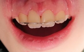
On extraoral examination of the patient numerous irregularities were observed in the number, position, and shape of teeth. Missing permanent teeth were discovered (only teeth 16, 14, 11, 21, 24, 26, 33, 43, 44, 46 were present) and persistent milk teeth (53, 52, 63, 73-83) (fig. 3-4). Teeth 11 and 21 were abnormally positioned and widely spaced, leading to the formation of a wide diastema (fig. 5-6). Conical shape of teeth 11, 21, 14, 24 was observed (fig. 3-6). Attrition (increased abrasion of teeth) and multiple missing permanent teeth (lack of support on lateral teeth) led to lowered occlusion (fig. 7-8). Pantomographic X-ray showed abnormalities in the structure of [...]

którzy są subskrybentami naszego portalu.
i ciesz się dostępem do bazy merytorycznej wiedzy!