Skuteczne leczenie zębów z różnymi rodzajami zmian endo-perio
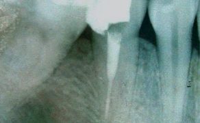
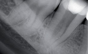
Local anesthesia (two 1.8 mL carpules of 2% lidocaine with 1:100,000 epinephrine) was then administered labially and palatally. A mucoperiosteal flap was raised mesially to the upper left canine tooth, and two vertical releasing incisions were formed in the anterior palatal area opposite teeth #21 and #23. A horizontal incision was formed from the left maxillary central incisor to the left maxillary canine. After flap reflection, a sling suture was placed in the tissue flap to secure it with the premolar tooth on the opposite side of the maxillary arch to aid the surgeon in improving visual and operative access by eliminating the need to manually retract the flap in the palatal area (fig. 3c) [8]. Cortical bone was absent on the mesial side of tooth #22. The root surface was covered with black calculus and the area was obliterated with granulation tissues. After removing the granulation tissues and calculus from the root surface with ultrasonic tips (figs. 3c, d), bone graft material (Puros Particulate Allograft; Zimmer Biomet Dental) mixed with saline was placed into the bony defect with a plastic instrument, and a resorbable barrier membrane (CopiOs membrane, Zimmer Biomet Dental) was placed above the bone graft. The flap was then repositioned and sutured with 4-0 Vicryl thread (Ethicon Inc., Somerville, NJ, USA). The patient was prescribed Augmentin (amoxicillin (875 mg) and clavulanic acid (125 mg)) (GlaxoSmithKline (Ireland) Ltd.) 1 g twice daily for 5 days and ibuprofen (600 mg) orally every 6 h for 2 days. Follow-up of the patient [...]

którzy są subskrybentami naszego portalu.
i ciesz się dostępem do bazy merytorycznej wiedzy!