Wpływ terapii fotodynamicznej (PDT) na biofilm Enterococcus faecalis w pierwotnych i wtórnych zakażeniach doświadczalnych kanału korzeniowego
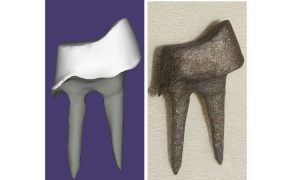
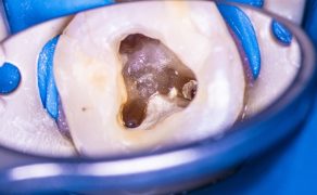
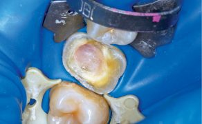
Standard access cavities were prepared and the precise tooth length was determined by inserting an ISO 10 K-file (VDW®, Munich) into the canal until the file was visible at the apical foramen. The tip of the file was then placed exactly at the apical foramen of the tooth to measure the tooth length. Working length (WL) was set 1 mm short of tooth length. Root canals were prepared using ProTaper instruments (Dentsply®) according to the manufacturer’s instructions in combination with an Endo IT Professional (VDW®) at 250 rpm. During root canal preparation a 3% sodium hypochlorite solution was used for irrigation [25]. Root canal preparation was performed using ProTaper shaping files S1 and S2 and finishing files F1 and F2. Apical preparation was performed terminating with ProTaper F2 file at WL. A ProTaper F2 gutta-percha point was set at WL and the apical region was covered with composite using Optibond FL (Kerr Corporation) and CeramX mono (Dentsply®) for an apical seal. Teeth were embedded in methacrylate (Techovit 4070TM, Haereus Kulzer®) and then decoronated using a diamond rotary cutting instrument (6830 L.314.014, Gebr. Brasseler GmbH & Co. KG) at 40.000 rpm at the cementum-enamel junction. After a final irrigation with 3% sodium hypochlorite and 5% sodium thiosulfate, the teeth were subsequently autoclaved at 134°C for 18 minutes. To verify that the root canals were free of microorganisms, samples were taken from the root canals using sterile ProTaper F2 paper points. Paper points incubated in the root canal for [...]

którzy są subskrybentami naszego portalu.
i ciesz się dostępem do bazy merytorycznej wiedzy!